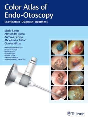
Color Atlas of Endo-Otoscopy - Examination - Diagnosis - Treatment
| Verlag | Thieme |
| Auflage | 2017 |
| Seiten | 348 |
| Format | 23,5 x 31,7 x 2,2 cm |
| Hardback (Thread Stitching) | |
| Gewicht | 1566 g |
| Artikeltyp | Englisches Buch |
| ISBN-10 | 3132415235 |
| EAN | 9783132415232 |
| Bestell-Nr | 13241523A |
A powerful guide to the primary diagnosis of disorders of the external auditory canal, tympanic membrane, middle ear, temporal bone, and skull base
Despite the many advances in diagnostic technologies and imaging modalities in recent years, otoscopy remains the first diagnostic option in the diagnosis of otologic disease.
This is an easy-to-consult book for residents and specialists, featuring brilliant diagnostic images from the newest generation of endoscopic otoscopes. Written by a renowned team of experts with 30 years of experience, this book helps readers obtain proficiency in otoscopy and in the interpretation of findings. Readers will learn what clinical consequences the diagnoses may have through case examples and treatment suggestions.
Key Features:
- Richly illustrated with over 1000 mostly full-color photographs and many radiological studies
- Shows a vast range of common and rare pathologies that can be visualized and assessed via en do-otoscopy
- Juxtaposes, when appropriate, the clinical picture, radiological diagnosis, and intraoperative findings with the endo-otoscopic findings of the patient
- In each chapter, a surgical summary lists various approaches that may be used to optimally plan treatment of the patient
- A special final chapter covers the assessment of postsurgical findings as seen in otoscopy, so as to distinguish between normal healing and changes that may require further intervention
Color Atlas of Endo-Otoscopy, produced with the support of Mario Sanna Foundation, is certain to become a valuable tool for all physicians involved in the care of patients with ear ailments.
Inhaltsverzeichnis:
1. Methods of Otoscopy
2. The Normal Tympanic Membrane
3. Diseases Affecting the External Auditory Canal
4. Otitis Media
5. Cholesterol Granuloma
6. Atelectasis, Adhesive Otitis Media
7. Noncholesteatomatous Chronic Otitis Media
8. Chronic Suppurative Otitis Media with Cholesteatoma
9. Congenital Cholesteatoma of the Middle Ear
10. Petrous Bone Cholesteatoma
11. Temporal Bone Paragangliomas
12. Rare Retrotympanic Masses
13. Meningoencephalic Herniation
14. Postsurgical Conditions

