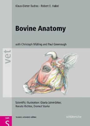
Bovine Anatomy
| Verlag | Schlütersche |
| Auflage | 2011 |
| Seiten | 176 |
| Format | 25,2 x 1,8 x 35,0 cm |
| Gewicht | 1333 g |
| Artikeltyp | Englisches Buch |
| ISBN-10 | 3899930525 |
| EAN | 9783899930528 |
| Bestell-Nr | 89993052A |
Die zweite englische Auflage dieses erfolgreichen Lehrbuches ist nun auch nach dem bewährten Konzept der "Budras-Atlanten" durch namhafte Experten aus der Anatomie und der klinischen Medizin um die klinisch-funktionelle Anatomie erweitert.
"This is a much-needed textbook-atlas that depicts bovine anatomy. It is appropriately organized such that it can easily be the single book that veterinarians refer to when an anatomic question needs to be answered about this species. It is most definitely worth the price."
JAVMA - Journal of the American Veterinary Medical Association
This expanded second edition provides detailed information on the structure, function, and clinical application of all bovine body systems and their interaction in the live animal.
Bovine Anatomy provides the reader with detailed information on the structure, function, and clinical application of all bovine body systems and their interaction in the live animal. The expanded second edition now includes clinical anatomy and retains the topographical and systems based methods of anatomy used in the first edition. The topographic anatomy is accompanied by systematic illustrations of the bones, joints, muscles, organs, blood vessels, nerves, and lymph nodes for each body system. There are also tables containing detailed information on the muscles, lymph nodes, and peripheral nerves.
The authors pay particular attention to the histology, growth, and function of the bovine hoof. In addition to the gross anatomy of the udder, its development, histology, and function are described and illustrated. One chapter is devoted to the pathology, pathogenesis, and molecular biology of bovine spongiform encephalopathy, scrapie of sheep and goats, and chronic wasting diseas e.
Each page of text is followed by a full page of colour illustrations. The Second Edition also contains more than 70 new diagrams and clinical photographs. The book has long been acknowledged as a valuable reference for study and revision, and this new edition is an essential resource for practitioners and students alike.
Published by Schluetersche, Germany and distributed by Manson Publishing.

