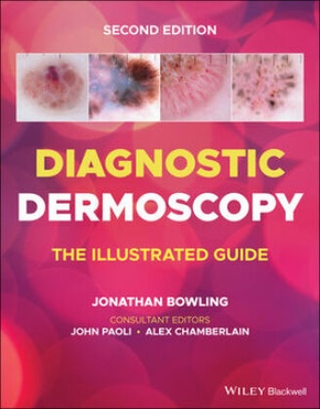
Diagnostic Dermoscopy - The Illustrated Guide
| Verlag | Wiley & Sons |
| Auflage | 2022 |
| Seiten | 336 |
| Format | 22,7 x 1,6 x 27,7 cm |
| Gewicht | 946 g |
| Artikeltyp | Englisches Buch |
| EAN | 9781118930489 |
| Bestell-Nr | 11893048UA |
DIAGNOSTIC DERMOSCOPY
DIAGNOSTIC DERMOSCOPY
Diagnostic Dermoscopy delivers a focused, practical and user-friendly guide to diagnosing the most frequently seen presentations of skin lesions practitioners encounter in clinic. The new edition offers a comprehensive collection of strategies to find and diagnose skin cancers at an earlier point in their evolution, and to increase confidence in diagnosing benign lesions to reduce both patient morbidity and mortality.
The many full-colour illustrations included within are standardised to aid in recognition, while the adjoining dermoscopy images focus on critical diagnostic features which, when combined with the provided clinical information, are sufficient for specific diagnoses or narrow differential diagnoses. The new edition is formatted to aid teledermoscopy education, including many illustrated cases for reference. Diagnostic Dermoscopy offers:
_ A comprehensive overview of melanocytic lesions, melanoma pr esentations, and non-melanocytic lesions
_ Thorough explorations of basal cell carcinoma and keratinocyte dysplasia
_ Practical discussions of several special sites, including acral, nail, facial, and scalp lesions, as well as trichoscopy
_ In-depth examinations of vascular lesions, inflammoscopy for general dermatology, and entomodermoscopy
_ Detailed illustrations of genetic and iatrogenic lesions
_ Tips from international experts in melanoma diagnosis
Diagnostic Dermoscopy is the ideal resource for dermatologists, primary care physicians, plastic surgeons and allied health practitioners involved in skin lesion and melanoma diagnosis.
Dermoscopy UK
Further information on dermoscopy education, and online courses can be found at www.dermoscopy.co.uk
Inhaltsverzeichnis:
1. Introduction - banner colour: white
2. Melanocytic lesions - banner colour: brown
3. Melanoma chapter - banner colour: black
4. Non-melanocytic lesions - banner colour: purple
5. Basal cell carcinoma chapter - banner colour: dark blue
6. Keratinocyte dysplasia chapter - banner colour: light blue
7. Special sites: Acral lesions chapter - banner colour: dark green
Special sites: Nail lesions chapter - banner colour: mid green
Special sites: Facial lesions chapter - banner colour: pale green
Special sites: Scalp lesions chapter - banner colour: bright green
Special sites: Trichoscopy - banner colour: light bright green
8. Vascular lesions - banner colour: blood red
9. Inflammoscopy (general dermatology) - banner colour: pink
11. Entodermoscopy chapter - banner colour: orange
10. Genetic lesions - banner colour: yellow
12. Iatrogenic lesions (miscellaneous) - bann er colour: grey
